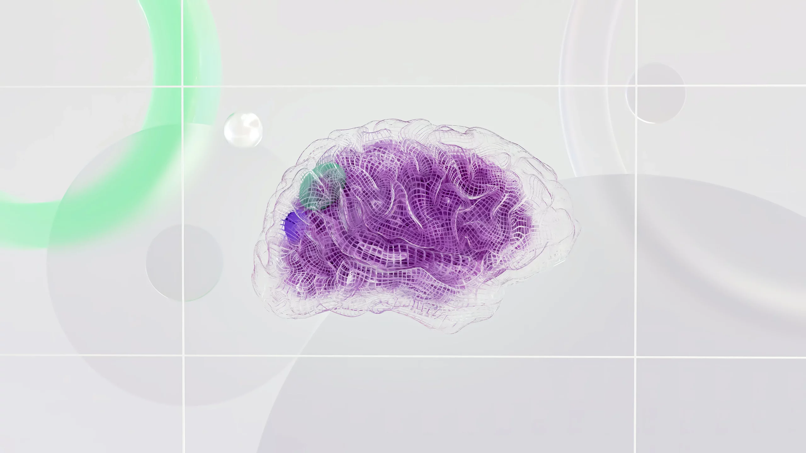
The Neurobiology of Obsessive Compulsive Disorder
Leave Your Info
What The Research Tells us About Obsessive Compulsive Disorder
Studies of neurophysiological disorders, like OCD and other neuro-based disorders have focused on specific brain regions, primarily on the orbitofrontal cortex (OFC) and basal ganglia, in its pathophysiology. Furthermore, recent neurobiological studies of OCD have found a close correlation between clinical symptoms, cognitive function, and brain function. Researches have observed patterns of high activities in the OFC in individuals with OCD.
The Neurobiology of Obsessive Compulsive Disorder & Attention
The concept of attention consists of three elements: information processing, attention span, and selective attention. Many researchers have tested for deficits in information processing and attention span using cognitive tests. Although studies have demonstrated decreased information processing speed, there is little evidence for a marked impairment of these functions in OCD. On the other hand, in terms of selective attention, some researchers hypothesize that patients with OCD have difficulty switching attention. Additionally, results from an fMRI study was conducted focusing on task-switching and and found that patients with OCD had significantly higher error rates in trials and reduced activation in prefrontal regions of the brain. Thus, neuro-imaging studies may provide evidence of attention-processing disturbances in OCD.
The Neurobiology of Obsessive Compulsive Disorder & Executive Function
Another benefit of understanding the neurobiology of obsessive compulsive disorder is its applicability to managing issues with executive function. Executive function is the higher cognitive function that controls and manages other cognitive processes. This includes planning, cognitive flexibility, rule-taking, changing sets, and problem-solving abilities. Because the concept of executive function may be plausible in explaining the persistent and inflexible thinking and behavior of OCD, many researchers have looked at executive function in OCD. Similarly, The Wisconsin Card Sorting Test (WCST) is suitable for testing overall variability. From this research, it was observed that individuals with OCD scored lower that those without OCD.
However, there is research that suggests that this is a result of a lack of cognitive flexibility. An fMRI study showed significantly stronger left prefrontal cortex activation in patients with OCD compared with normal controls when running the word generation test. Therefore, it seems that executive impairment may be a likely explanation for behaviors such as inadequate decision making or lack of flexibility in changing behavior commonly seen in obsessive compuslive disorder.
The Neurobiology of Obsessive Compulsive Disorder & Working Memory
Working memory is one of the higher cognitive functions involved in mediating integration, processing, removal, and retrieval of information. It differs from the general memory function in that it includes active monitoring and information manipulation. In this regard, some researchers have found impaired working memory in obsessive compulsive disorder using the Tower of London test. An fMRI study showed that patients with OCD performed poorly at the highest levels of difficulty and engaged in the same pool of brain regions as matched healthy control regions in the midbrain, prefrontal cortex regions and peaks, particularly related to working memory
How Neurobiology of Obsessive Compulsive Disorder Can Inform Today's Treatment
While there is certainly a specific biological basis for OCD, such as the OCD loop, the complex involvement of different neural networks may underlie its clinical diversity and lead to deviations in responses to treatment. At present, we can derive a certain amount of information from neurobiological studies, but there are many questions that need to be clarified.

Studies of neurophysiological disorders, like OCD and other neuro-based disorders have focused on specific brain regions, primarily on the orbitofrontal cortex (OFC) and basal ganglia, in its pathophysiology. Furthermore, recent neurobiological studies of OCD have found a close correlation between clinical symptoms, cognitive function, and brain function. Researches have observed patterns of high activities in the OFC in individuals with OCD.
Neurobiology of Obsessive Compulsive Disorder: Treatment Options
There are current treatment options available for obsessive compulsive disorder and we are confident that the services at Mind by Design can help you on your journey to OCD recovery. With various options for interventions and modalities, your treatment plan will be tailored to your unique needs. Read about our OCD treatment here:
Rebecca is a certified clinical anxiety treatment specialist and provides treatment for OCD, Phobias and Panic Disorder using Exposure Therapy, Virtual Reality Therapy and Mindful-Based CBT. Learn more by clicking here.
Finding the Right Help for OCD
How do I get started as a new client?
New Clients can reach out to us directly via call, text or email here:
What is your cancellation policy?
We ask that clients provide at least 24 hours notice in the event that they need to cancel to avoid the 50% cancellation fee. we understand that life happens and do our best to be flexible & reschedule.
Does my insurance cover my visits?
We provide”Courtesy Billing” for clients who are using the Out-of-network insurance benefits.
Our Insurance Page shares a small blurb about Why We Left Insurance Panels
Do you offer traditional talk therapy?
of course! though we have some unconventional therapy approaches, we are rooted in evidenced based practices. Talk therapy is a major player in the therapy room! See What we Treat and Integrative Services for more information
Is Online Therapy As Effective As In-Person Therapy?
Online therapy is essentially face-to-face counseling, just conducted remotely. Studies show that teletherapy is as effective as traditional counseling. Professional organizations and state governments recognize its benefits and have set regulations for it. However, like any therapy, its success in achieving your goals isn’t guaranteed. It’s important to discuss with your therapist whether teletherapy is working for you.
Can I Change Therapists If I'm Not Happy?
Yes, you can switch therapists to another provider within the practice, or we can provide you a referral if preferred. We want to ensure that your time and effort are well spent, and that you are getting the relief you need, that’s why we work collaboratively with each other in the practice, as well as outside therapists who we know and trust.
How Do I Know If Therapy Is Helping?
You should feel like you’re making progress. Signs it’s working include:
Feeling comfortable talking to your therapist
Your therapist respects boundaries
You’re moving towards your goals
You feel listened to
You’re doing better in life
Your self-esteem is getting better
Is Online Therapy Easy to Use for Non-Tech-Savvy People?
Yes, it’s pretty simple to access sessions. You’ll need basic internet skills, such as opening and visiting the patient link sent to you via email. It’s similar to video chatting like Facetime or Zoom. We can also walk you through it on the phone the first time to ensure a strong connection
What Questions Should I Ask My New Therapist?
Feel free to ask anything. Some good questions are:
- How often will we meet?
- What do you specialize in?
- What experience do you have with my issue?
- What outcomes can I expect?
- How will I know I’m progressing?
- How long do you usually work with clients?
- How will we set my treatment goals?
How Should I Prepare for My First Session?
Showing up is all that you need to do! But if you really want to get the most out of session, it could help to take some time to think about what you want from therapy. It helps to write down your goals, questions you have or things that you feel are important to share.
What is the difference between associate therapists & fully licensed therapists?
Our Qualifications:
Our founder, Rebecca Sidoti, is a highly qualified, state-licensed therapist and supervisor with extensive training in anxiety related disorders and innovative treatment such as Ketamine Therapy. Mind by Design Counseling adheres to standards set by the our governing counseling boards.
To see each providers credentials, training and licenses, visit our “Meet the Therapists” Page to learn more.
- LAC/LSW are therapists who may practice clinical work under the supervision of a fully licensed therapist.
- LPC/LCSW are therapists who have completed the necessary clinical hours post-graduation under supervision and can practice clinical work independently.
What Geographic Areas Are Served?
Currently, we serve clients in New Jersey and are expanding to other states as telehealth laws evolve. While telehealth offers the convenience of attending sessions from anywhere, state laws require clients to be in-state during their session.
Is Virtual Counseling Suitable for Everyone?
Online therapy might not be as effective for individuals with chronic suicidal thoughts, severe trauma, significant mental health history, or those recently in intensive care. Such cases often benefit more from traditional, in-person counseling. We’ll help you decide if our online services are right for you during your intake and evaluation.
What Equipment is Needed for Online Therapy?
To join a session, log in using the credentials we provide. No downloads are needed. Our platform, compatible with both individual and group sessions, requires:
A computer or mobile device with a webcam and internet access.
We’ll help you test your setup before your first appointment to ensure a reliable connection. iOS users should use the Safari browser for mobile and tablet sessions.
What Questions Will Therapists Ask Me?
It depends on your goals. Expect questions about your thoughts, feelings, relationships, work, school, and health. They’ll ask to understand your therapy goals.
How Do You Keep Client Information Secure?
Security and Confidentiality of Sessions:
Your privacy is crucial to us. We use TherapyNotes, a HIPAA-compliant platform, ensuring secure and confidential teletherapy sessions. This platform’s security features include encrypted video connections, secure data transfers, and encrypted databases, ensuring your information is safe at all times.
What is VRT used for?
we use VRT to support Exposure Therapy, a long standing traditional therapy modality to treat phobias, anxiety and stress. we send a headset directly to your home so you can access VRT from anywhere.
VRT not only helps with exposure therapy for phobias, but is great for ADHD, mindfulness, PTSD and social anxiety.
References:
- Martinot JL, Allilaire JF, Mazoyer BM et al. Obsessive-compulsive disorder: A clinical, neuropsychological and positron emission tomography study. Acta Psychiatr. Scand. 1990; 82: 233–242.
- Schmidtke K, Schorb A, Winkelmann G, Hohagen F. Cognitive frontal lobe dysfunction in obsessive-compulsive disorder. Biol. Psychiatry 1998; 43: 666–673.
- Nakao T, Nakagawa A, Yoshiura T et al. A functional MRI comparison of patients with obsessive-compulsive disorder and normal controls during a Chinese character Stroop task. Psychiatry Res. 2005; 139: 101–114.
- Gu BM, Park JY, Kang DH et al. Neural correlates of cognitive inflexibility during task-switching in obsessive-compulsive disorder. Brain 2008; 131: 155–164.
- Head D, Bolton D, Hymas N. Deficit in cognitive shifting ability in patients with obsessive-compulsive disorder. Biol. Psychiatry 1989; 25: 929–937.
- Abbruzzese M, Ferri S, Scarone S. Wisconsin Card Sorting Test performance in obsessive-compulsive disorder: No evidence for involvement of dorsolateral prefrontal cortex. Psychiatry Res. 1995; 58: 37–43.
- Gross-Isseroff R, Sasson Y, Voet H et al. Alternation learning in obsessive-compulsive disorder. Biol. Psychiatry 1996; 39: 733–738.
- Lucey JV, Burness CE, Costa DC et al. Wisconsin Card Sorting Task (WCST): Errors and cerebral blood flow in obsessive-compulsive disorder (OCD). Br. J. Med. Psychol. 1997; 70: 403–411.
- Cavedini P, Ferri S, Scarone S, Bellodi L. Frontal lobe dysfunction in obsessive-compulsive disorder and major depression: A clinical-neuropsychological study. Psychiatry Res. 1998; 78: 21–28.
- Pujol J, Torres L, Deus J et al. Functional magnetic resonance imaging study of frontal lobe activation during word generation in obsessive-compulsive disorder. Biol. Psychiatry 1999; 45: 891–897.
- Purcell R, Maruff P, Kyrios M, Pantelis C. Neuropsychological deficits in obsessive-compulsive disorder: A comparison with unipolar depression, panic disorder, and normal controls. Arch. Gen. Psychiatry 1998; 55: 415–423.
- Mataix-Cols D, Junqué C, Sànchez-Turet M et al. Neuropsychological functioning in a subclinical obsessive-compulsive sample. Biol. Psychiatry 1999; 45: 898–904.
- van der Wee NJ, Ramsey NF, Jansma JM et al. Spatial working memory deficits in obsessive compulsive disorder are associated with excessive engagement of the medial frontal cortex. Neuroimage 2003; 20: 2271–2280.
- Nakao T, Nakagawa A, Nakatani E et al. Working memory dysfunction in obsessive-compulsive disorder: A neuropsychological and functional MRI study. J. Psychiatr. Res. 2009; 43: 784–791.
- Kang DH, Kwon JS, Kim JJ et al. Brain glucose metabolic changes associated with neuropsychological improvements after 4 months of treatment in patients with obsessive-compulsive disorder. Acta Psychiatr. Scand. 2003; 107: 291–297.
- van der Wee NJ, Ramsey NF, van Megen HJ et al. Spatial working memory in obsessive-compulsive disorder improves with clinical response: A functional MRI study. Eur. Neuropsychopharmacol. 2007; 17: 16–23.


